Best Anatomy of the Ear Surgeon in Ahmedabad
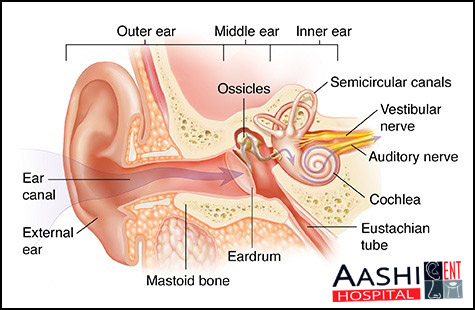
- The ear allows you to hear and helps to maintain your sense of balance. It is divided into three parts – the outer, middle and inner ear.
- The outer ear consists of the earlobe or pinna, and the ear canal. It has a protective lining of hairs and glands that produce wax, which traps dust and stops small objects from entering the ear canal. The outer ear directs sound into the middle ear.
- The middle ear is an air-filled cavity that contains three small bones, or ossicles. The bones are called the malleus, incus and stapes. A thin membrane, the eardrum, separates the middle ear from the ear canal. When sound travels down the ear canal, the eardrum vibrates. This vibration is amplified by the ossicles and then passed to the inner ear.

- The middle ear is connected to the nose by the eustachian tube, and to another air-filled cavity behind the ear called the mastoid. The eustachian tube is normally closed, but opens when you yawn or swallow.
- The inner ear is delicate and complex. It contains the cochlea, which changes sound vibrations into a nerve signal, and the vestibule and semicircular canals, which maintain your sense of balance and position.

